|
I recently read this article and I was intrigued about how nutrients in red meat can affect the risk of you developing atherosclerosis.
Various experiments have been undertaken involving mice to investigate further into what nutrients present in red meat have an effect on atherosclerosis. The team of scientists from Cleveland Clinic Learner Research institute in Ohio fed the mice a diet high in carnitine which is a substance found in red meat. They found that whilst feeding the mice this diet the incidences of atherosclerosis increased, however, to compare their results they also fed mice with supressed gut flora the exact same diet and found that there was no increase in atherosclerosis. Based on previous knowledge we already know that certain bacteria in the intestine use the substance carnitine as an energy source, and when broken down, a waste product known as trimethylamine (TMA) is produced. This TMA is then converted at the liver into TMAO, which is the substance that brings about this increased risk of atherosclerosis. From the mice experiment, they found that a carnitine-rich diet boosted certain bacteria types and as a result it increased the level of TMAO. It is this TMAO that increases the uptake of cholesterol likely to build-up and cause plaques and the TMAO will also prevent the cholesterol from being broken down by macrophages. Therefore this can lead to atherosclerosis developing. To back this evidence up, a series of further tests underwent only to find that meat-eaters produced higher levels of TMAO than vegetarians after they were both fed carnitine, which suggests that those meat-eaters, who eat meat on a regular basis, had more bacteria which produce TMA in the intestine. QUICK SUMMARY - These experiments suggest that eating red meat regularly could affect our arteries leading to an increased risk of developing atherosclerosis. Red meat contains carnitine which boosts the number of bacteria which use carnitine to produce energy. These specific bacteria produce TMA as a waste product when breaking the carnitine down, which will be converted into TMAO in the liver. It is this TMAO which is linked to an increased risk of development of atherosclerosis. https://www.newscientist.com/article/dn23352-red-meat-boosts-gut-bugs-that-raise-heart-disease-risk/
1 Comment
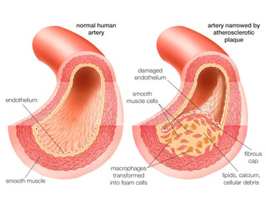
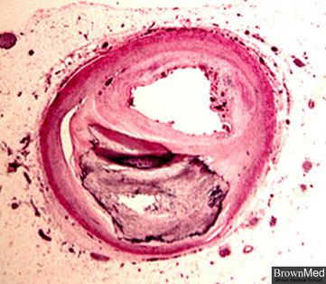
Introduction Atherosclerosis is a build-up of plaques (fatty deposits) on the inside of the arteries. A plaque can build up until it restricts the flow of blood through the artery which increases blood pressure and there is a chance of the plaque completely blocking the artery to a point when no blood can pass through. Plaque tends to build up in the arteries surrounding the heart (coronary arteries) and neck (carotid arteries). The problem with restricted blood flow is that organs become damaged and stop working properly which could be fatal. Also if a plaque ruptures it will lead to the formation of a blood clot which could potentially block off the blood supply to the heart or brain resulting in a heart attack or stroke. In this way atherosclerosis is linked to peripheral arterial disease where the blood supply to the legs is blocked and coronary heart disease where the coronary arteries become clogged with plaques. How does atherosclerosis develop? It begins with damage to the endothelial lining of blood vessels which may be caused by high blood pressure or the chemicals in tobacco smoke. Atherosclerosis occurs in arteries rather than veins because the endothelial lining is more likely to get damaged when the blood is flowing under high pressure. Once the damage to the lining has occurred, the body responds and signals for white blood cells to arrive at the site. These cells accumulate chemicals in the blood such as cholesterol which is one of the main constituents of plaque. As a result of accumulation of these chemicals an atherome will form on the arterial lining. Calcium salts and fibrous tissue also build up and surround the plaque which hardens it, therefore this area of the artery will lose some flexibility. This build-up of plaque will narrow the lumen of the artery which increases blood pressure. Higher blood pressure will cause further damage elsewhere leading to more build-up of plaques. As a result blood pressure will get even higher which will cause more problems. Effect on health? Atherosclerosis leads to a raised blood pressure - a higher pressure will damage smaller delicate blood vessels. For example: blood vessels leading to the kidneys the eye and the brain will become damaged as these vessels are not strong enough to take the force of an increased pressure of blood. Aneurysms – as the plaque continues to accumulate, blood can build up behind the clump which puts the artery wall under more pressure than usual which causes it to weaken. This may lead to the artery splitting open causing internal bleeding. Heart disease - angina is a common example of heart disease involving blockage of the coronary arteries. As plaques build-up in the arteries surrounding the heart, blood flow becomes more and more restricted. As a result the heart is pumping on a restricted supply of oxygen meaning it may have to respire anaerobically at times. This process brings about a pain in the chest which may spread into the arms and often causes breathlessness. Another common example of heart disease would be the heart attack also known as myocardial infarction. This is more severe than angina as it involves complete blockage of a coronary artery resulting in a section of the heart lacking an oxygen supply. A heart attack occurs as a result of a part of the heart being starved of oxygen. Strokes – this involves the blood supply to the brain being temporarily cut off. It can be caused by blockage in the artery due to plaque build-up or blood clots. What increases the risk of developing atherosclerosis? The risks of developing atherosclerosis can be increased by several factors some of which cannot be modified as you were born with it and some which can be modified by a change in lifestyle.
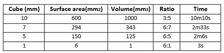
An experiment to investigate the effect of surface area : volume ratio on the diffusion rate.2/9/2016 The objectives of this experiment was to calculate the surface area to volume to ratio of pieces of agar and determine what effect this ratio had on the rate of diffusion. Equipment:
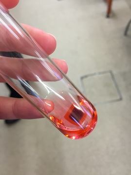
What happens when the agar is placed in the HCl? As time goes on the colour of the agar block becomes increasingly orange as it loses its pink colour. The reason for this is because of the hydrochloric acid diffusing into the block and making it lose its colour. The acid travels from an area of high concentration of HCl (in the test tube) to an area of low concentration of HCl (the agar cube) along a concentration gradient and the point at which the original colour has completely gone is when an even distribution of particles has been achieved. This experiment was designed to then see how this diffusion process occurred in different sized cubes and to investigate the difference I looked at the diffusion rate, i.e. how long it took for the pink colour to disappear. My prediction would be that the rate of diffusion would speed up as the surface area to volume ratio was increased, i.e. the cube was made smaller.
 Conclusion
From these results I can see that the surface area to volume ratio ad an effect on how fast diffusion occurred. As the agar cubes were cut smaller the ratio of surface area to volume became larger and this affected the diffusion rate by making it faster. The results show a clear trend that supports my knowledge on how the diffusion rate speeds up when there is a larger surface area to volume ratio. The time decreased which highlights how diffusion occurred quicker as there was more surface area in relation to its volume over which the HCl could diffuse over. Another reason for this increase in rate was due to the fact that the HCl particles had to travel longer distances in those agar cubes which had a larger volume in comparison to its surface area. Overall my results support the idea that as the surface area to volume ratio increases so does the rate of diffusion. This experiment relates to real life because single-celled organisms use diffusion as their means of getting the substances they need and expelling those they don’t. Diffusion works efficiently for single-celled organisms such as amoeba due to a large surface area to volume ratio as there is a relatively large surface area over which substances can diffuse into or out of the organism. This is demonstrated in my experiment as the smaller cubes with a larger surface area to volume ratio resemble single-celled organisms. My results also support the idea that large organisms cannot use diffusion as their means of transporting substances in and out, as shown by the 10mm by 10mm cube which took 10 minutes and 10 seconds for the HCl to diffuse all the way through. Diffusion would not be efficient enough for larger organisms as the surface area is not large enough for substances to diffuse over, as a result they require a complex transport system. This experiment does have its limitations as the organism’s cell is represented by a lifeless agar cube. It lacks a semi-permeable membrane and only allows for simple diffusion unlike real-life membranes. Cell surface membranes are much more sophisticated as they consist of a combination of phospholipids, carbohydrates and carrier/channel proteins. As well as this the shape of the agar cube does not resemble an organism’s cell. Apart from these limitations this experiment is a good way of demonstrating the effect of surface area to volume ratio on the rate of diffusion. I carried out this experiment to investigate what affect placing a layer of an onion on a microscope slide in sucrose solution would have in comparison to placing one in water to act as the control.
Conclusion
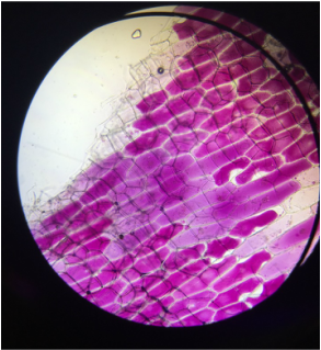
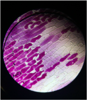
From these two images taken from the microscope it is clear to see that the onion cells that were in water remained turgid as the cytoplasm is firmly pushing against its cell wall, (despite a few which appear flaccid). The reason for this would be due to water moving into the cells by osmosis down a concentration gradient. On the other hand, the onion cells placed in sucrose solution looked different to those in water. From the image I can see that some of the cells have started to become flaccid as the cells lose water. The reason for this loss of water from the cell is because there is a higher concentration of water in the onion cell compared to its surroundings meaning water moves from an area of higher water potential to an area of low water potential by osmosis. Some of the cells in the microscopic image have clearly undergone plasmolysis meaning the cytoplasm no longer firmly pushes against the cell wall but instead has pulled away from it creating irregular spaces in the cell. The second image demonstrates excessive loss of water due to osmosis and the cells irregular shape highlight how this can be a problem if the cells lose too much water as they are no longer turgid and will not be as strong as it is the turgidity of the cells that provides the plant with support. |
Ciara Branagan
Archives
October 2016
|
 RSS Feed
