|
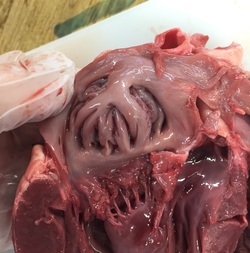
The structure of the respiratory system in a fish... Salmon is an example of a bony fish with high oxygen demand. Due to having an impermeable outer scaly surface, exchange of gases must be completed through a respiratory system. Gills control gas exchange in fish. They are vital organs situated at either side of the fish’s head and each gill consists of a row of gill filaments. Each of these filaments contains lamellae which are essentially discs containing capillaries resulting in a rich blood supply. This ultimately maintains the steep concentration gradient so that diffusion of gases is efficient and the exchange process is as quick as possible. Another adaptation of the fish would be the number of these gill filaments. There are a lot of these filaments which increases the surface area over which gas exchange can occur which will increase the rate of diffusion further. These gills are located within the gill cavity and are covered by a protective bony flap known as the operculum. This structure is important in maintaining a constant flow of water over the gills to allow for continued extraction of oxygen. To maximise gas exchange, as well as a large surface area, rich blood supply and thin walls, fish have what’s known as a countercurrrent exchange system. This means that the blood in the gill filaments is flowing in the opposite direction to the movement of water over the gills. This increases the concentration gradient, allowing oxygen absorption to be more efficient. The process... To dissect this fish I used a scalpel, tweezers and scissors and I placed the fish head on a tray. To ensure my skin didn’t come into contact with the fish I wore gloves whilst dissecting. Firstly I removed the operculum to reveal the gill filaments inside. The structure of the several gill arches was clear to see and I was also able to identify where the blood vessels were located in the gill arch. The gill filaments were arranged in rows and were densely packed, which explains why gas exchange is so efficient in fish due to a large surface area. I removed one of the gill arches using a scalpel to take a closer look at the structure and relate it back to diagrams of the gases passing through the gaps in between the filaments. I then cut off a section of a filament and mounted it onto a slide and placed it under a light microscope. This revealed a close up view of a filament and once the image was focused I was able to see small black dots which could have potentially been blood clots in the filament. Equipment:
Method:
Safety: There are certain hazards involved in the dissection of the insect including the risk of cutting oneself with the scissors or parts of the insect squirting into the eye. To overcome these hazards, you could wear safety goggles and ensure, when dissecting, to cut away from oneself. What I would expect to see based on knowledge and research? From the insect I would expect to see spiracles along the thorax and abdomen. These are the site of entry and exit of the respiratory gases. It is these spiracular valves that control gas exchange as well as controlling water loss. Despite having fairly high metabolic rates, many insects have periods when their spiracles are closed, temporarily reducing gas exchange. This adaptation is involved in preventing excessive water loss. There is also the argument stating the spiracles can’t always be open to prevent the tracheae tissues from being exposed to high levels of oxygen for too long which could potentially cause damage. However the insect must let in some oxygen and remove carbon dioxide to prevent toxic gas build up. To enable this movement of gases, the sphincters surrounding the spiracles contract and relax to open and close. The tracheae also make up the insects tracheal system. They are the largest tubes of the respiratory system (up to 1mm). The function of the tracheae is to carry air directly into the body for gas exchange with the cells. The tracheae are supported by chitin spirals – holds the tubes open to prevent them collapsing. These tubes were identifiable under the light microscope and it was clear that there is a huge network of tracheae tubes so that as much gas can be exchanged as possible. This is particularly important for the very active insects with high metabolic rates. The tracheoles are the thinner tubes which the tracheae divide into and they are the main site of gas exchange. These tubes are much smaller (0.6-0.8μm) meaning they are harder to see under the light microscope. This was one of the limitations of this experiment, I believe it could have been overcome by analysing the insect under a stronger microscope. What I saw? From my dissection of the locust I was able to identify the spiracles on the thorax and abdomen of the insect and through further inspection I was able to see the network of tracheae under the light microscope. This experiment has helped me to visualise the structure of the insect’s tracheal system and it has given me a better understanding of the role of each component to the respiratory system of the insect. Bibliography Edexcel AS/A level Biology B1 Pearson textbook www.nature.com/nature/journal/v433/n7025/abs/nature03106.html- 18/01/16 |
Ciara Branagan
Archives
October 2016
|


 RSS Feed
