|
I carried out this experiment to investigate what affect placing a layer of an onion on a microscope slide in sucrose solution would have in comparison to placing one in water to act as the control.
Conclusion
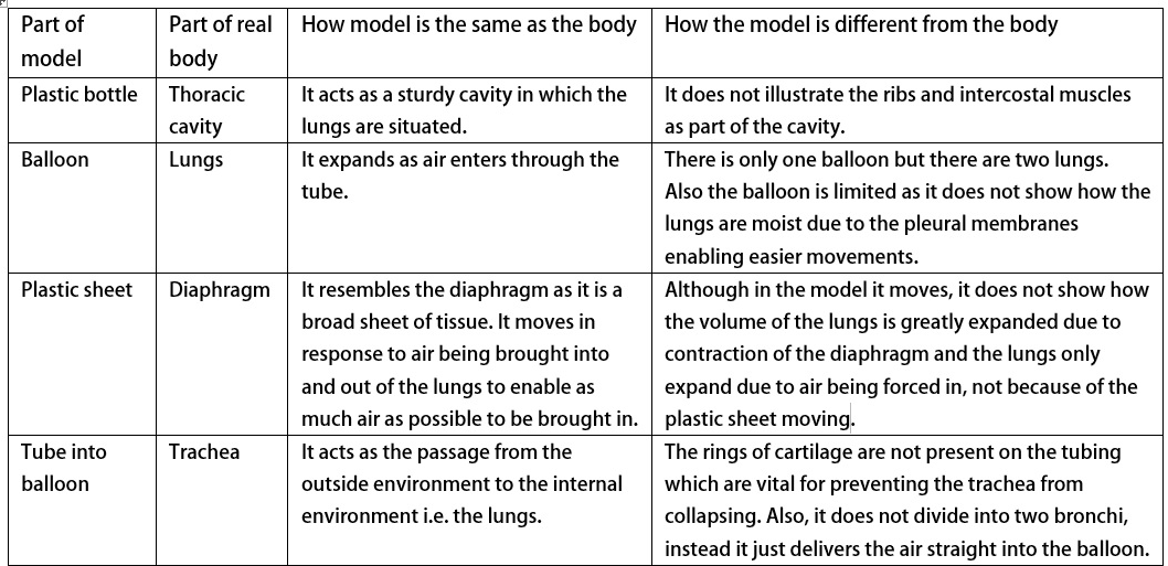
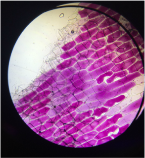
From these two images taken from the microscope it is clear to see that the onion cells that were in water remained turgid as the cytoplasm is firmly pushing against its cell wall, (despite a few which appear flaccid). The reason for this would be due to water moving into the cells by osmosis down a concentration gradient. On the other hand, the onion cells placed in sucrose solution looked different to those in water. From the image I can see that some of the cells have started to become flaccid as the cells lose water. The reason for this loss of water from the cell is because there is a higher concentration of water in the onion cell compared to its surroundings meaning water moves from an area of higher water potential to an area of low water potential by osmosis. Some of the cells in the microscopic image have clearly undergone plasmolysis meaning the cytoplasm no longer firmly pushes against the cell wall but instead has pulled away from it creating irregular spaces in the cell. The second image demonstrates excessive loss of water due to osmosis and the cells irregular shape highlight how this can be a problem if the cells lose too much water as they are no longer turgid and will not be as strong as it is the turgidity of the cells that provides the plant with support.
1 Comment
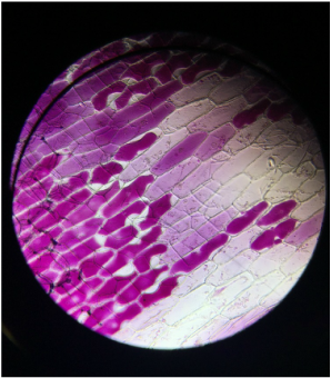
The structure of the respiratory system in a fish... Salmon is an example of a bony fish with high oxygen demand. Due to having an impermeable outer scaly surface, exchange of gases must be completed through a respiratory system. Gills control gas exchange in fish. They are vital organs situated at either side of the fish’s head and each gill consists of a row of gill filaments. Each of these filaments contains lamellae which are essentially discs containing capillaries resulting in a rich blood supply. This ultimately maintains the steep concentration gradient so that diffusion of gases is efficient and the exchange process is as quick as possible. Another adaptation of the fish would be the number of these gill filaments. There are a lot of these filaments which increases the surface area over which gas exchange can occur which will increase the rate of diffusion further. These gills are located within the gill cavity and are covered by a protective bony flap known as the operculum. This structure is important in maintaining a constant flow of water over the gills to allow for continued extraction of oxygen. To maximise gas exchange, as well as a large surface area, rich blood supply and thin walls, fish have what’s known as a countercurrrent exchange system. This means that the blood in the gill filaments is flowing in the opposite direction to the movement of water over the gills. This increases the concentration gradient, allowing oxygen absorption to be more efficient. The process... To dissect this fish I used a scalpel, tweezers and scissors and I placed the fish head on a tray. To ensure my skin didn’t come into contact with the fish I wore gloves whilst dissecting. Firstly I removed the operculum to reveal the gill filaments inside. The structure of the several gill arches was clear to see and I was also able to identify where the blood vessels were located in the gill arch. The gill filaments were arranged in rows and were densely packed, which explains why gas exchange is so efficient in fish due to a large surface area. I removed one of the gill arches using a scalpel to take a closer look at the structure and relate it back to diagrams of the gases passing through the gaps in between the filaments. I then cut off a section of a filament and mounted it onto a slide and placed it under a light microscope. This revealed a close up view of a filament and once the image was focused I was able to see small black dots which could have potentially been blood clots in the filament. Equipment:
Method:
Safety: There are certain hazards involved in the dissection of the insect including the risk of cutting oneself with the scissors or parts of the insect squirting into the eye. To overcome these hazards, you could wear safety goggles and ensure, when dissecting, to cut away from oneself. What I would expect to see based on knowledge and research? From the insect I would expect to see spiracles along the thorax and abdomen. These are the site of entry and exit of the respiratory gases. It is these spiracular valves that control gas exchange as well as controlling water loss. Despite having fairly high metabolic rates, many insects have periods when their spiracles are closed, temporarily reducing gas exchange. This adaptation is involved in preventing excessive water loss. There is also the argument stating the spiracles can’t always be open to prevent the tracheae tissues from being exposed to high levels of oxygen for too long which could potentially cause damage. However the insect must let in some oxygen and remove carbon dioxide to prevent toxic gas build up. To enable this movement of gases, the sphincters surrounding the spiracles contract and relax to open and close. The tracheae also make up the insects tracheal system. They are the largest tubes of the respiratory system (up to 1mm). The function of the tracheae is to carry air directly into the body for gas exchange with the cells. The tracheae are supported by chitin spirals – holds the tubes open to prevent them collapsing. These tubes were identifiable under the light microscope and it was clear that there is a huge network of tracheae tubes so that as much gas can be exchanged as possible. This is particularly important for the very active insects with high metabolic rates. The tracheoles are the thinner tubes which the tracheae divide into and they are the main site of gas exchange. These tubes are much smaller (0.6-0.8μm) meaning they are harder to see under the light microscope. This was one of the limitations of this experiment, I believe it could have been overcome by analysing the insect under a stronger microscope. What I saw? From my dissection of the locust I was able to identify the spiracles on the thorax and abdomen of the insect and through further inspection I was able to see the network of tracheae under the light microscope. This experiment has helped me to visualise the structure of the insect’s tracheal system and it has given me a better understanding of the role of each component to the respiratory system of the insect. Bibliography Edexcel AS/A level Biology B1 Pearson textbook www.nature.com/nature/journal/v433/n7025/abs/nature03106.html- 18/01/16 Bluefin tuna are amongst the most active fish meaning they need efficient respiratory and circulatory systems to supply their needs. A Bluefin swims with its mouth open, forcing water past its gills in a process called ram ventilation. Its gills have up to 30 times more surface area than those of other fish. Respiratory systems of southern Bluefin tunas are adapted to their high oxygen demand. Bluefin tunas are obligate ram ventilators: they drive water through their mouth (buccal cavity), then over the gills, while swimming. It is this movement of oxygenated water over the gills that allows the fish to stay alive. The Bluefin does not require a separate pump to produce a stream of water over the gills. However, when you compare this feature to other species of fish, separate pumps are present. Bluefin don’t need this pump system because it isn’t efficient enough to provide the volume of oxygen needed. This means that tuna must keep swimming to allow the water to pass over the gills to ultimately to prevent suffocation. The oxygen need and oxygen uptake of the southern Bluefin tuna are related. As the tuna increases its energy need by swimming faster, water flows into the mouth and over the gills more quickly, increasing the oxygen uptake. Tunas have highly specialized gills, with a surface area larger than that of other marine environment organisms. This increased surface area allows more oxygen to be in contact with the respiratory surface and therefore diffusion occurs quicker. Also it has a very thin gas-exchange membrane allowing the oxygen to diffuse a short distance across the respiratory surface to get to the blood. This allows the tuna to take oxygenated blood into the circulatory system more quickly. As well as this, the tuna’s uptake of oxygen from the water is a lot more efficient in comparison to other species of fish. The Bluefin tunas rate of utilization of oxygen has been recorded as high as 60% whilst other species typically utilize below this at roughly 27-50%. This is due to a combination of the large surface area of the gills and the short distance the oxygen has to travel. This means that oxygen can be supplied quicker enabling the highly metabolic organism to maintain an optimum temperature and energy for continuous swimming. This then allows the fish to continue its search for food to grow and reproduce and ultimately stay alive Ultimately the adaptations of the respiratory system compliment the Bluefin’s circulatory system which contains more haemoglobin, meaning the blood has a greater oxygen capacity. Comparing the Bluefin tuna's respiratory system with the human respiratory system When comparing the Bluefin’s respiratory system to that of humans there is a clear difference in the efficiency of the process. The tuna requires a much more efficient system because it gains its oxygen from a denser medium, water. Uptake of the oxygen from water must be effective because water contains less oxygen than air itself. Although the efficiency differs, they are the similar in the sense that their features greatly increase the surface area allowing for more efficient gas exchange by diffusion. Also both systems consist of thin, moist membranes to allow the movement of dissolved gases to pass into the blood. Another obvious difference would be the location of the respiratory organs, as in fish they are situated to the sides of the organism whilst in a human the lungs occupy the upper torso. A huge difference would be that the tuna requires movement to pass the water over the gills (ram ventilation), whereas in a human it is the contraction of internal muscles which alters the pressure in the lungs forcing air in or out. This highlights the need for the external environment to provide the movement for oxygen to be taken in for fish, in comparison to the internal environment of the thoracic cavity bringing about oxygen intake. Overall the two systems share the same purpose but have different adaptations due to differing environments. https://en.wikipedia.org/wiki/Southern_bluefin_tuna
http://link.springer.com/article http://ngm.nationalgeographic.com/2014/03/bluefin-tuna/superfish-interactive https://www.researchgate.net/publication/226448900_Cardiovascular_and_respiratory_physiology_of_tuna_Adaptations_for_support_of_exceptionally_high_metabolic_rates http://www.s-cool.co.uk/a-level/biology/gas-exchange/revise-it/gas-exchange-in-fish
 Method:
Aseptic techniques to follow:
My results table: From this table I have decided to discard a number of the results because they are anomalies. The anomalous results are due to human error combined with misuse of the colorimeter. Prior to the experiment my group didn’t know that the colorimeter gave out two different readings; one of absorbance of light and the other transmission of light. This meant that when I recorded our results, it was hard to spot a trend due to a clear difference between those which were transmission (i.e. negative) and those of absorbance (between 0 and 1). Therefore, to overcome this problem, I inverted those negative values and plotted them on the graph. On top of this, other anomalies were generated which I believe could be easily improved upon. Firstly, I would take more care in ensuring the liquid broth was agitated and cloudy before taking a sample from it. This could be done by swirling the broth to allow the yeast cells to be more evenly distributed in the liquid. After removing the significant anomalies, I generated a new table and then plotted a graph of absorbance of light against time in hours.   This graph demonstrates a general trend of upward growth. To begin with the growth is more rapid but as the time increases the gradient of the curve becomes less steep. Evaluating the results there is a trend in the growth of the yeast, despite a number of anomalies, there is still general upward growth. To begin with the yeast cells are adapting to their environment in the growth medium and have not yet reached their maximum rate of reproduction. This is not shown on the graph due to the fact that the earliest recording we took was after 18 hours of incubation. I believe that if we took earlier readings and replotted the graph, the gradient would have resembled the typical lag phase of growth. This would be a key area for improvement. However, because this was not the case, the graph I created began with the exponential growth phase. As the curve continues at a steep gradient, the exponential phase of growth is very clear to see. This phase is where yeast reproduction is at its highest due to availability of nutrients and an optimum temperature. The gradient eventually becomes less steep demonstrating the stationary phase where the number of yeast cells produced is not increasing. This is shown on the graph as the gradient begins to level off. This may be due to competition over space or nutrients as it reaches its carrying capacity. 
Aseptic technique is to carry out laboratory procedures with sterile equipment and to prevent microbial contamination. Equipment:
The aim of this experiment was to move a sample of the monoculture from the test tube into the nutrient agar medium whilst avoiding contamination from foreign microorganisms. Many techniques were carried out to prevent contamination from occurring. The method:
This method requires the aseptic techniques to be carried out carefully. Another aseptic technique would be that during the experiment it is best not to place the equipment down as that could cause further contamination and the longer the instruments are exposed to the air the more bacteria are likely to build up and cause microbial contamination. We recently looked into the basics of synthetic biology and I learnt about how living organisms can be so easily manipulated in the lab by interfering with the natural processes such as protein synthesis. I found it interesting what synthetic biologists have predicted will be possible in the near future such as creating bullet-proof flies. The possibilities are endless and it's weird to think how synthetic biology is the gateway to creating new species with abilities that were previously thought to be beyond our reach. The advancement in technology and equipment has enabled progress in synthetics. Through understanding the way in which transcription and translation works, scientists have the ability to manipulate parts of the genetic code and assemble it in any way. This creates new sequences which will lead to coding for different proteins, therefore it will be able to carry out different functions.
|
Ciara Branagan
Archives
October 2016
|


 RSS Feed
